One Diagnosis, Many Presentations: Why Skin of Color Changes the Clinical Picture

Photo: Internet Archive Book Images, No restrictions, via Wikimedia Commons
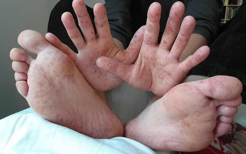
For decades, the reference image in dermatology education was the same: a pale, pink lesion on fair skin. If a condition looked different on brown or Black skin — and many do — the textbook often did not say so. That gap in representation has had measurable consequences in exam rooms across the United States, where patients with skin of color face longer delays to diagnosis, more advanced disease at detection, and worse outcomes for conditions that are highly treatable when caught early.
A growing movement within the specialty is working to change that — and patients have an important role to play in the effort.
When the Textbook Image Fails
Consider melanoma, the deadliest common skin cancer. On lighter skin, melanoma frequently appears as an asymmetrical dark spot with irregular borders — the 'ABCDE' criteria many Americans know from public health campaigns. On darker skin, melanoma is more likely to arise in less obvious locations: the palms, the soles of the feet, under a nail, or on mucous membranes. Acral lentiginous melanoma, the subtype most associated with deeper skin tones, may look brown, black, or even lack striking pigmentation altogether.
The result is a stark disparity. While melanoma incidence is lower among Black and Hispanic Americans, five-year survival is persistently worse — a difference researchers attribute largely to later detection rather than biology alone.
Other common conditions tell a similar story:
- Eczema on darker skin often appears violet, ashen, or gray rather than red, and post-inflammatory hyperpigmentation can linger for months after the itch resolves.
- Psoriasis may look purple or brownish with less visible scale, leading patients and even clinicians to mistake it for other conditions.
- Keloidal scarring and conditions like central centrifugal cicatricial alopecia disproportionately affect people with textured hair and darker skin, yet receive a fraction of the research funding devoted to more common presentations.
The Equity Problem in the Numbers
The consequences compound across the health system. Studies have documented that patients with skin of color are more likely to be diagnosed with advanced skin cancer, more likely to report that a clinician told them 'Black people don't get skin cancer,' and less likely to receive timely referrals. Teledermatology added a new wrinkle: early studies found that remote image-based diagnosis performed worse when photographs were taken without proper lighting or exposure calibrated for darker skin.
Meanwhile, the dermatology workforce itself remains one of the least diverse in American medicine. Representation matters not only for trust and communication, but because physicians who have trained in communities of color are more likely to recognize conditions as they actually present.
How the Specialty Is Responding
Change is underway on several fronts:
Curriculum reform. Medical schools and dermatology residencies are overhauling image libraries and didactic materials to include multiple skin tones for every condition, a shift accelerated by open-access resources built specifically for skin of color.
Inclusive imaging technology. Engineers are recalibrating dermatoscopes, smartphone attachments, and AI diagnostic tools to render pigmented skin accurately, addressing the lighting and exposure biases baked into older systems.
Dedicated clinical centers. Skin of color clinics at academic medical centers across the country are expanding access to specialists experienced in textured hair, pigmentary disorders, and conditions with atypical presentations.
Research investment. Advocacy from professional societies and patient groups has pushed funders and journals to require diverse study populations and representative clinical photography.
What Patients Can Do Right Now
Until training and technology fully catch up, self-advocacy remains a powerful tool:
- Photograph anything new or changing. A dated photo series of a spot on the palm, sole, or nail bed gives your dermatologist a timeline.
- Describe what you see, not what you think you should see. 'This patch turned darker and grayish' is more useful than 'it's not really red.'
- Ask directly. 'How does this condition typically look on my skin tone?' is a fair and important question.
- Seek a second opinion. If a concerning lesion is dismissed without examination, a board-certified dermatologist — ideally one with experience in skin of color — can offer another evaluation.
- Know your risk. A family history of melanoma, a changing nail streak, or a non-healing sore on the sole warrants a prompt exam regardless of skin tone.
Accurate diagnosis should not depend on what a patient's skin looks like in a textbook. Closing the gap requires reform in classrooms, laboratories, and clinics — and patients who arrive informed, documented, and unafraid to ask for the answers they deserve.